医疗城 · 设备实验室 · 第 21 台,共 69 台
经皮肾镜碎石系统 (PCNL)
PCNL System
BMEssentia 医疗城设备实验室中的经皮肾镜碎石系统 (PCNL)。以可交互 3D 打开设备,逐个部件拆解,了解其工作原理,然后在 BMEssentia 平台上系统学习,并向 Essentia 提问。
本页的设备实验室内容为英文,与医疗城中的内容一致。
- 10 个部件
- 设备实验室中有 2 种配置
- 又称: Percutaneous Nephrolithotomy, Nephroscope
设备实验室
PCNL System
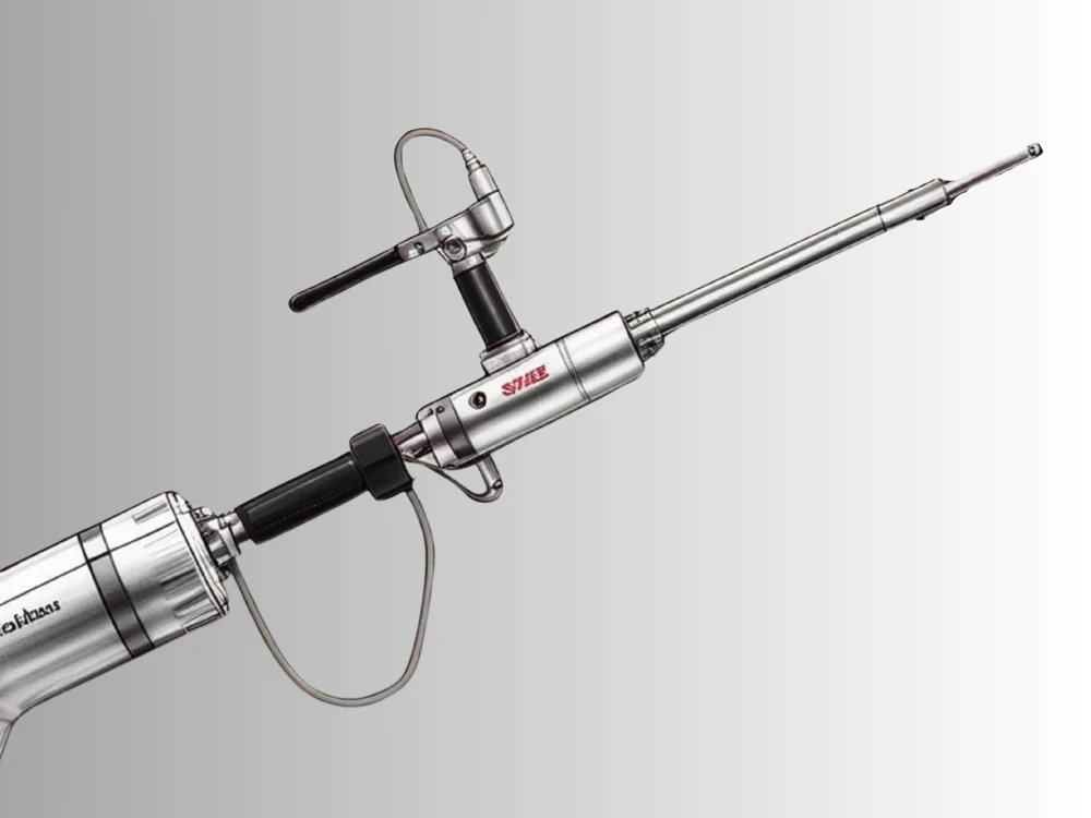
A handpiece in which compressed air drives a small projectile down a short barrel into the back of a solid probe, transmitting one impact per cycle at a rate the operator sets. The probe passes down the working channel of a 24 French rigid nephroscope, which sits inside the access sheath. There is no lumen in the probe and no aspiration anywhere on the system, so fragments are picked out individually with a grasper passed down the same channel, and the instrument is withdrawn and reinserted for each retrieval.
配置
设备实验室中的各个版本。
Pneumatic ballistic lithotripter, solid probe
A handpiece in which compressed air drives a small projectile down a short barrel into the back of a solid probe, transmitting one impact per cycle at a rate the operator sets. The probe passes down the working channel of a 24 French rigid nephroscope, which sits inside the access sheath. There is no lumen in the probe and no aspiration anywhere on the system, so fragments are picked out individually with a grasper passed down the same channel, and the instrument is withdrawn and reinserted for each retrieval.
Ultrasonic lithotripter, hollow probe with integrated suction
A piezoelectric stack behind a horn, driving a hollow titanium probe at its own resonance, with the probe lumen connected to a suction pump through a trap. The probe passes down the working channel of a 26 French rigid nephroscope, larger than the ballistic configuration needs because a hollow probe with a usable lumen cannot be made as slender as a solid one. Fragments are drawn up the lumen as they are produced, so the instrument stays in the sheath for most of the case and the fragments arrive in the trap rather than on a grasper.
工作原理
在设备实验室中探索。
The pressure nobody sets and nobody reads
Leave every instrument alone and move only the bag height. Read the cavity pressure on the bench schematic and the inflow on the flow readout at one metre, then at half a metre, then at a quarter. Then put the bag back and change the ureteric bore instead.
Two French of instrument, and a factor of four
Set the sheath to 30 French and leave it there for the whole experiment. Put a 24 French nephroscope in it and read the pressure. Change only the nephroscope size to 26 French and read it again.
What a second outflow subtracts, and where it crosses zero
This experiment needs the configuration that has a lumen. Set the suction to nothing and read the cavity pressure. Raise it to 200 mL/min and read it again.
A temperature rise with no upper bound
Again the configuration with a lumen. Set the drive power and read the temperature rise of the stream leaving the probe at 200 mL/min, then at 50, then at 20. Then leave the flow alone and change the power instead.
A push that is not zero, and one that is exactly zero
Two experiments side by side, one on each configuration, both of them about a single quantity: the impulse delivered to the stone.
部件
逐个部件讲解。
Percutaneous access sheath and its bore
A short rigid tube with a bevelled tip and a flared collar at the outer end, thirty French across and nine and a half millimetres in the bore, standing in a tract about a hundred and twenty millimetres long. It is the only structure holding the route open, and it is the outflow path of the entire procedure.
Rigid nephroscope, offset eyepiece and working channel
A straight rigid instrument with an eyepiece offset to one side, a light post, an irrigation tap and a working channel running the length of it. It is the object that does everything: it carries the optics, the light, the irrigant and the probe.
Rod-lens telescope and the light post
A sealed optical train running the length of the nephroscope: a train of glass rods with thin air gaps between them, an objective at the distal end, an eyepiece at the proximal end, and a fibre bundle alongside it ending at a post on the side of the instrument. It is the only reason anybody can work inside the sheath at all.
The clearance between the sheath and the instrument
Not an object at all. A ring-shaped space, four tenths of a millimetre to three quarters of a millimetre wide depending on what is standing in the sheath, running the length of the tract.
Irrigation bags, the hanger and the height that drives the inflow
Two or three bags of warmed irrigant on a hanger near the top of the mast, one running and the others waiting, with a scale on the mast recording how high the hanger is. The height is the drive.
Irrigation set, drip chamber, warmer and roller clamp
Two metres of clear tubing, four millimetres in the bore, with a spike at one end, a drip chamber below it, a warming section, a roller clamp and a connector for the nephroscope tap. It looks like the most substantial part of the inflow path and it is very nearly the least important part of it.
The scope irrigation channel and its resistance
80 millimetres across and three hundred millimetres long, running inside the nephroscope from the irrigation tap to the tip. It is the shortest part of the inflow path, the smallest object in this lesson, and the largest single resistance anywhere in the device.
Drape pocket, collection bag and the weighed recovery
A pocket moulded into the drape below the access site, a length of tubing from it to a graduated bag on the frame, and a set of scales. It collects what comes back up the annulus, and the number it produces is the only measured quantity in the whole fluid argument.
Cold light source, lamp module and fibre cable
A box on the trolley with a lamp module in it, a fan, an intensity control, and a socket for a fibre cable that runs to the post on the nephroscope. It is called a cold light source because the heat stays in the box rather than because the light arriving at the tip is cool.
Camera head, coupler and the video processor
A camera head clipped to the eyepiece through a coupler, a cable to a processor box, and the processing that turns a sensor output into a picture. It is the only sensing chain on this device that reports anything continuously, and what it reports is light.