المدينة الطبية · مختبر الأجهزة · الجهاز 44 من 69
قياس ضغط المريء
Oesophageal Manometry
قياس ضغط المريء في مختبر الأجهزة في المدينة الطبية من BMEssentia. افتحه بتقنية ثلاثية الأبعاد تفاعلية، وفكّكه قطعةً قطعة، وتعرّف إلى طريقة عمله، ثم تعلّمه على منصة BMEssentia واسأل إسنشيا عنه.
محتوى مختبر الأجهزة في هذه الصفحة باللغة الإنجليزية، كما هو داخل المدينة الطبية.
- عدد الأجزاء: 10
- عدد التكوينات في مختبر الأجهزة: 2
- يُعرف أيضًا باسم: Esophageal Manometry, High-Resolution Manometry
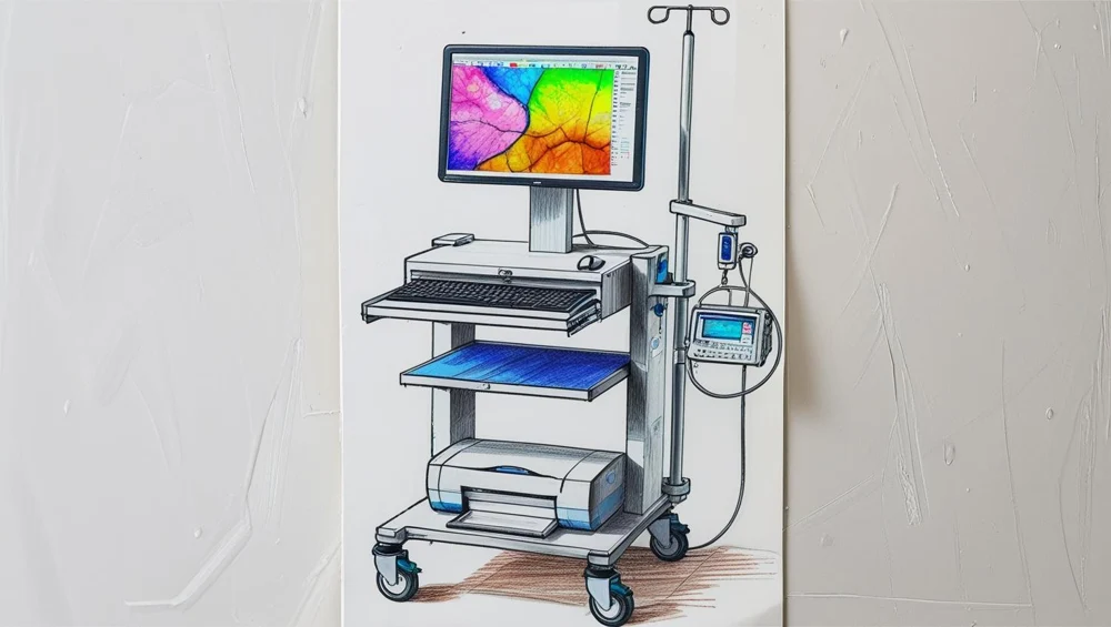
مختبر الأجهزة
Oesophageal Manometry
An extruded multi-lumen catheter with one side-hole per lumen, spaced along the assembly, each lumen carried out of the patient to its own external transducer on a bank beside the workstation. A capillary pump driven from a pressurised reservoir pushes a small steady flow down every lumen and out of every hole, so the pressure at each transducer is the pressure needed to keep that flow leaving that hole. A sleeve sensor spans the lower sphincter, because a sphincter three centimetres long cannot be relied on to contain any hole at this spacing.
التكوينات
نسخ الجهاز في مختبر الأجهزة.
Water-perfused side-holes with external transducers
An extruded multi-lumen catheter with one side-hole per lumen, spaced along the assembly, each lumen carried out of the patient to its own external transducer on a bank beside the workstation. A capillary pump driven from a pressurised reservoir pushes a small steady flow down every lumen and out of every hole, so the pressure at each transducer is the pressure needed to keep that flow leaving that hole. A sleeve sensor spans the lower sphincter, because a sphincter three centimetres long cannot be relied on to contain any hole at this spacing.
Solid-state circumferential sensors at one centimetre
Thirty-six pressure-sensing elements built into the catheter itself at one centimetre intervals across the same span, each element made of sectors distributed around the circumference whose outputs are averaged into one number. There is no perfusate, no lumen and no external transducer, so there is nothing to infuse, nothing to flush and nothing to level. The sleeve reading over the lower sphincter is computed from the sensors that span it rather than provided by a physical sleeve, which is only possible because there are enough of them.
كيف يعمل
استكشفه في مختبر الأجهزة.
What one column of the plot loses, and why it goes as the square
Generate a single travelling band of stated length, sample it at one spacing, then at the other, and read the highest value the reconstruction offers across the body at a single instant. Compare it against the highest value the field actually has at that instant. Then sweep the phase of the grid against the band and keep the worst case.
The same sampling, two thirds as much lost
Take the same band and, instead of the maximum, compute the double integral above the twenty millimetre contour over the axial window and over the record - which is what the study actually quotes, and whose unit says so: millimetres of mercury times seconds times centimetres. Compute it from the samples with a trapezoidal rule and again from the closed form of the field, at both spacings.
A finer spacing is worse at speed, and then it is not
Time the crossing of the thirty millimetre contour on two neighbouring sensors and divide the spacing by the difference between the two instants. Do it for a single pair on each configuration. Then do it the way the device does it, averaging every adjacent pair that lies inside the tubular body, and compare the two answers.
A rate limit is not a bandwidth, and the obvious answer is wrong
Drive one channel with a slow rise and then with a fast one of the same amplitude, and read what comes out of each. A body contraction reaching a hundred millimetres of mercury in two seconds is fifty a second. A pharyngeal transient reaching two hundred in a hundred and fifty milliseconds is thirteen hundred and thirty-three a second.
A gap shorter than one spacing has somewhere to hide
Put a break of a stated length into the contractile band on the bench model, then slide the break along the axis in small steps and, at every position, count how many sensors lie inside it. Do it with a break longer than the spacing and again with one shorter.
An average round a closed circle removes something exactly
Write the pressure round one axial station as a mean plus a single harmonic - a constant plus an amplitude times the cosine of the angle minus a phase - and read it two ways. First as the average of twelve sectors distributed evenly round the circumference. Then as one unidirectional hole facing in one direction.
الأجزاء
قطعةً قطعة.
Catheter assembly, its size and its depth markings
An extruded tube a little over four millimetres across and about a metre long, graduated in centimetres from the tip, with a moulded hub at the outside end and a serial number printed on the shaft. It is the only object in this device that decides where every sensing station is, and the marks along it are the only link between a channel number and a place inside a person.
Transnasal route, anaesthesia and the gag
A nostril, a gel, a swallow of water and a person who has to tolerate a tube through the back of the throat for half an hour while being asked to swallow on command. No part of the airway or the pharynx is drawn in this model; the assembly enters at the nostril and the model stops there.
Nasal fixation and the catheter clip
A strip of tape at the nose, a clip on the shoulder of the gown, and a loop of slack between the two. Nothing about it is electronic and nothing about it is optional, because the horizontal axis of every plot in the study is a geometry that this is the only thing holding.
The swallow protocol and its timing
Ten swallows, one at a time, on command, separated by twenty to thirty seconds, with a period of quiet before them and the whole sequence written on a card beside the workstation. It is the least mechanical component in the device and it is the one that decides how many independent looks at the field the study gets.
The measured bolus and its syringe
A small syringe of water, a stated volume, and a cup. Five millilitres per swallow is the usual figure.
The spatial pressure plot and the interpolation in it
A rectangle of colour with time across it and position up it, apparently a continuous picture of pressure everywhere along the tube at every instant. It is the single most persuasive object on this machine and almost none of it is data.
Finding the two sphincters and following them as they move
A pair of horizontal bands on the plot that are darker than the body between them, and a tool for saying where their edges are. The upper one sits near the top of the span and the lower one near the bottom, and both of them move while the study is running.
The sleeve reading and the relaxation window
A channel that does not report the pressure at a point. It reports the highest pressure anywhere along a stretch of the assembly, and on one configuration it is a physical object while on the other it is a calculation.
Contractile front velocity, from two sensors and a clock
A number in centimetres per second, obtained by asking two neighbouring stations when each of them crossed a contour and dividing the distance between them by the difference between the two instants. No sensor in the device measures a speed; this is entirely arithmetic done on two timestamps.
The contractile integral, over space and over time
The figure a manometry study actually quotes about a contraction, and it cannot be measured at a point by definition. It is pressure above a contour, summed over a length of the axis, summed over the time the contraction takes, and its unit says so: millimetres of mercury times seconds times centimetres.